Breaking News


Popular News


Contents

2D ultrasound imaging is a common diagnostic technique that uses high-frequency sound waves to create images of the inside of the body. This technology is widely used in medical settings to visualize the internal organs and tissues of patients.
Ultrasound technologists play a crucial role in operating the equipment and capturing the images. They must have a strong understanding of anatomy and physiology to correctly interpret the images and assist healthcare providers in making accurate diagnoses.
Some common uses of 2D ultrasound imaging include monitoring fetal development during pregnancy, diagnosing abdominal conditions, and guiding needle biopsies. The real-time nature of the images allows for immediate feedback and adjustments during procedures.
Overall, 2D ultrasound imaging continues to be a valuable tool in the field of healthcare, providing non-invasive and cost-effective ways to gather important medical information.
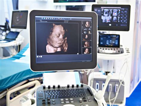
3D ultrasound technology has revolutionized the field of medical imaging by providing a more detailed view of the patient’s anatomy. This advanced imaging technique uses sound waves to create three-dimensional images of the body, allowing healthcare professionals to better visualize and diagnose medical conditions.
With 3D ultrasound technology, physicians can obtain a more accurate picture of internal organs, tissues, and structures. This can be especially useful in obstetrics, as it allows for a more detailed view of the developing fetus during pregnancy. The high-resolution images produced by 3D ultrasound can help detect abnormalities or potential health issues at an earlier stage.
One of the key benefits of 3D ultrasound technology is its ability to provide a clearer and more comprehensive view of the patient’s anatomy. This can lead to more accurate diagnoses and treatment plans, ultimately improving patient outcomes. Additionally, 3D ultrasound can reduce the need for additional imaging tests, minimizing cost and inconvenience for patients.
Overall, 3D ultrasound technology has significantly enhanced the capabilities of healthcare professionals in diagnosing and treating a wide range of medical conditions. As technology continues to advance, we can expect further improvements in 3D imaging quality and diagnostic accuracy, leading to even better patient care.
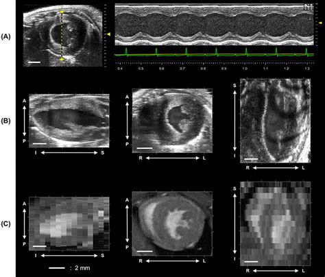
When it comes to advancements in ultrasound technology, 4D ultrasound has been a game-changer in the medical field. The ability to see a three-dimensional image in real-time provides healthcare professionals with a clearer view of the internal organs and structures of the body.
4D ultrasound technology offers improved visualization and accuracy compared to traditional 2D and 3D imaging. This has greatly enhanced the diagnosis and treatment of various medical conditions, allowing for more precise and efficient medical interventions.
With the use of ultrasound tech such as 4D imaging, medical professionals can better monitor fetal development during pregnancy, detect abnormalities in the fetus, and even perform guided procedures with higher precision.
Overall, 4D ultrasound advancements have revolutionized the way medical imaging is done, providing invaluable insights and improving patient outcomes in a wide range of medical specialties.
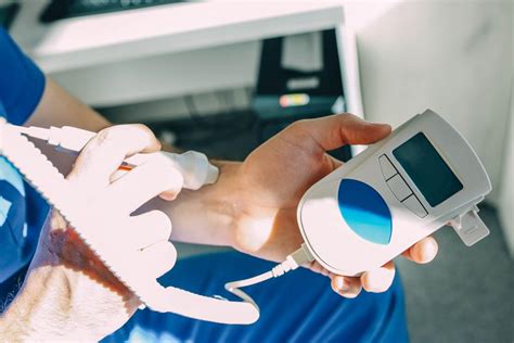
Doppler ultrasound technology plays a crucial role in the medical field, allowing healthcare professionals to assess blood flow in various parts of the body. By using sound waves to measure the direction and speed of blood flow, Doppler ultrasound can provide valuable information for diagnosing a range of conditions.
One application of Doppler ultrasound is in the assessment of cardiovascular health. By analyzing the blood flow in the heart and blood vessels, doctors can identify issues such as blockages or narrowing of arteries. This information helps in the early detection of cardiovascular diseases and allows for timely intervention.
Another important use of Doppler ultrasound is in obstetrics, where it is used to monitor blood flow in the placenta and fetus during pregnancy. By measuring the velocity of blood flow, healthcare providers can assess the health of the developing baby and detect any potential complications.
In addition, Doppler ultrasound technology is also used in the detection and monitoring of conditions such as deep vein thrombosis, where it can identify blood clots in the veins. By providing real-time images and data on blood flow, Doppler ultrasound helps in the accurate diagnosis and treatment of various medical conditions.
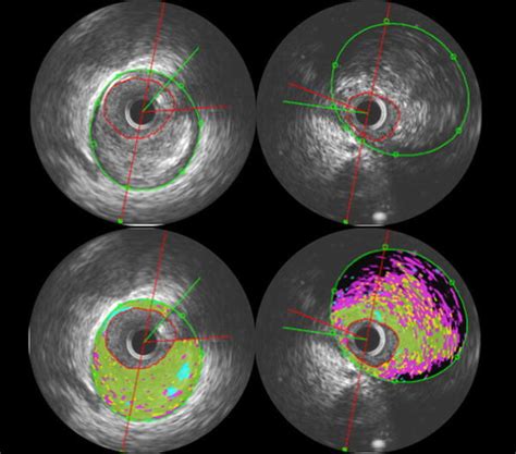
When it comes to medical imaging, ultrasound technology has revolutionized the way healthcare professionals are able to visualize the interior of the human body. Intravascular ultrasound techniques, in particular, have provided clinicians with a valuable tool for examining blood vessels from the inside.
Intravascular ultrasound, also known as IVUS, involves the use of a catheter with an attached ultrasound probe that is inserted into a blood vessel. This allows for real-time imaging of the vessel walls, providing detailed information about the structure and integrity of the vessel.
One of the key advantages of intravascular ultrasound techniques is the ability to assess areas that may not be easily visualized using other imaging modalities. This can be especially useful in guiding interventional procedures, such as angioplasty or stent placement, by providing precise information about the size and location of blockages within the vessel.
Overall, intravascular ultrasound techniques have significantly improved the ability of healthcare providers to diagnose and treat cardiovascular diseases, ultimately leading to better outcomes for patients.
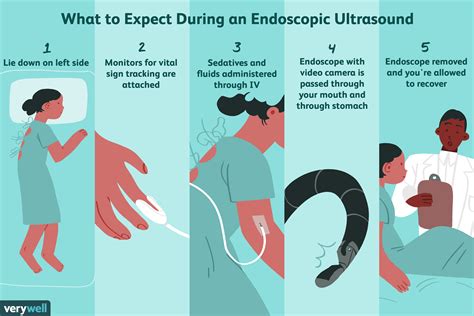
Endoscopic ultrasound procedures are a type of diagnostic imaging technique that combines endoscopy and ultrasound to visualize the organs and tissues within the body. This advanced technology enables healthcare providers to obtain detailed images of the gastrointestinal tract, pancreas, liver, and surrounding structures.
Using an endoscope equipped with an ultrasound transducer, the ultrasound tech can capture high-resolution images in real-time. This allows for a more precise and accurate diagnosis of various medical conditions, such as tumors, cysts, and abnormal growths.
| Benefits of Endoscopic Ultrasound Procedures |
|---|
| 1. Minimally invasive |
| 2. High diagnostic accuracy |
| 3. Ability to guide treatment decisions |
| 4. Low risk of complications |
Furthermore, endoscopic ultrasound procedures can also be used for therapeutic interventions, such as the drainage of fluid collections or the biopsy of suspicious lesions. This minimally invasive approach reduces the need for more invasive surgical procedures and shortens recovery times for patients.

There are several types of ultrasound technology, including 2D, 3D, and 4D ultrasounds. 2D ultrasounds create flat, two-dimensional images, while 3D ultrasounds create a three-dimensional image. 4D ultrasounds add the element of motion, providing a live video feed of the fetus.Ultrasound technology is generally considered safe for pregnant women when used in moderation. It does not use radiation like X-rays, but instead uses high-frequency sound waves to create images.Ultrasound technology is commonly used during pregnancy to monitor the development of the fetus. It is also used to diagnose medical conditions such as gallbladder disease, kidney stones, and heart abnormalities.Ultrasound technology works by sending high-frequency sound waves into the body and recording the echoes that bounce back. These echoes are then converted into images that can be viewed on a screen.While ultrasound technology is generally considered safe, there are some potential risks associated with prolonged or unnecessary exposure. It is important to use ultrasound technology only when medically necessary.3D ultrasounds create a three-dimensional image of the fetus, while 4D ultrasounds add the element of motion, providing a live video feed. 4D ultrasounds are often used for entertainment purposes, allowing parents to see their baby in real-time.Yes, ultrasound technology can be used for a variety of medical imaging purposes beyond pregnancy. It is commonly used to diagnose and monitor conditions such as muscle and joint injuries, organ abnormalities, and vascular disorders.